 |
|
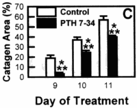 |
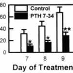
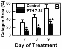
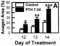
Figure 2. Effect ofPTH (7-34) on the hair cycle stage represented by macroscopic evaluation of percent area of back skin. (A) Two separate groups containing six to seven 24-d-old C57BL/6 mice were given either 1 µg of PTH (7-34) in 0.1 ml distilled water (n = 14) or vehicle alone (n = 13) twice daily intraperitoneally for 14 d. The skin color was evaluated as a macroscopic indicator of what hair cycle stage the hair follicles were in for the mice in each group on days 12, 13, and 14 of treatment. One control mouse died of unknown causes. (B) Two groups of seven 28-d-old anagen mice were given either 1 µg of PTH (7-34) (n = 14) or vehicle alone (n = 14) twice daily intraperitoneally for 9 d. The skin color of the mice from each group was evaluated on day 7, 8, and 9 of treatment. (C) Three groups of four to six 6- to 8-wk-old telogen mice were depilated to induce anagen. Starting on day 9 post depilation, when all mice had reached anagen, they were given either 1 µg PTH (7-34) (n = 15) or vehicle alone (n = 15) twice daily intraperitoneally for 10 d. The skin color of the mice from the treated and control groups were evaluated on day 9 and 10 and on the 11th day at the time of sacrifice. These panels compare the means from pooled data derived from two to three separate experiments of groups consisting of four to seven mice representing a total of 13 to 15 mice studied for each treatment day. The data and the means ± SEM and levels of significance were calculated using the Mann-Whitney U-test (***, p < 0.001; **, p < 0.01; *, p < 0.05). Each experiment by itself showed similar statistical significance.
cytes surrounding the hair follicle produce PTHrP (Merendino et al, 1986; Hayman et al, 1989), they do not possess a PTH/PTHrP receptor (Hanafin et al, 1995; Lee et al, 1995). It is not known whether the recently identified PTH2 receptor, which is found in the brain and pancreas, is present in the hair follicle or whether it recognizes PTH (7-34) (Usdin et al, 1995). Cultured human skin fibroblasts and the dermal papilla, however, express mRNA for the PTH/PTHrP receptor (Hanafin et al, 1995; Lee et al, 1995). Thus,
 |
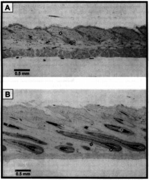 |
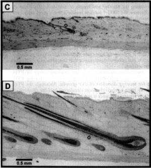
Figure 3. Effect of PTH (7-34) on hair follicle histology. (A) Acceleration of anagen development in 24-d-old, spontaneously cycling mice. On day 14 of treatment, the control mouse still showed mostly telogen follicles with condensed dermal papilla [dp’s] ( »). The skin is very thin. This is compared to the skin of the PTH (7—34)—treated mouse (B). The skin is not only much thicker (approximately 2 times) but also contained the characteristically large anagen hair bulbs with activated large dp’s ( »), that extend to the subcutis all producing melanin (3) and pigmented hair shafts (3). (C) Catagen inhibition/anagen prolongation in adolescent, depilation-induced anagen mice. Hair follicles from a mouse receiving vehicle alone showed the hair follicles in telogen on the 11th day of treatment (day 19 post depilation). Hair follicles from a mouse treated with PTH (7—34) showed the persistence of characteristic anagen VI hair follicles. (D) All pictures were taken with the same magnification (X 100) and represent comparable areas in the middle region of the back (A-D, scute bar, 0.5 mm).

